Introduction
Maxillary defects can be congenital or acquired, secondary to surgery due to neoplasms and life-threatening infections. Extensive surgical interventions result in significant anatomical and functional deficits leaving the patients debilitated, including challenges in mastication, speech, and aesthetics for rehabilitation. In 1978 Dr. Mohammed Aramany introduced the first published classification system for post-surgical maxillary defects according to relation of remaining natural teeth to the area of defect. This classification has been used for decades despite owing to emphasis of prosthodontic principles in guidance for prosthetic design. Understanding the need for support, retention, and stability in designing is crucial to achieving the objectives of prosthodontic care.1, 2, 3
Obturators can be classified into surgical obturators, interim obturators and definitive obturators. Obturators play a vital role in the rehabilitation of patients with maxillary defect. A definitive obturator is given after 3-6 months following surgery when no changes in soft tissue is expected. In addition to restoring lost soft and hard tissues, the obturator allows the patient to swallow, masticate, and speak nearly normally, while also creating a barrier between the nasal and oral cavities. Further benefits include its removable nature, facilitating clear visualization and early detection of any recurrent tumour. Additionally, the obturator contributes to improved facial appearance by providing support to facial tissues.4, 5, 6
This case report highlights the steps in fabrication of definitive obturator using semi-precision attachment (Rhein 83 OT Strategy) while still utilizing the basic principles of designing of cast partial denture in a patient with Armany Class 1 defect. The patient requirements of improved mastication, esthetics and phonetics were met. Prosthetic Rehabilitation of such patients help in restoring psychosocial well-being.
Case Presentation
A 59-year-old male patient reported to the Department of Prosthodontics with chief complaint of difficulty in eating food due nasal regurgitation. Systemic history revealed hypertension and diabetes since past 10 years. Dental history revealed that patient was a tobacco chewer for 10 years and has stopped since 3 years. Patient was apparently alright 3 years back but later reported with an ulcer and growth in left upper back region of mouth since 8 months. After thorough clinical, radiological and histological examination it was concluded to be a case of upper left alveolar carcinoma involving left hard palate, left buccal mucosa and gingivolabial sulcus. Hemi maxillectomy was done with adjuvant radiotherapy. Patient developed a surgical defect and was on nasogastric tube for a year after which an interim obturator was delivered. After 1 year the obturator was ill-fitting and patient had difficulty in eating with regurgitation of food and fluids into nasal.


Extraoral Examination showed facial asymmetry with depressed face on left side (Figure 1 A, B). TMJ was Normal. Reduced mouth opening of 22mm was noted. Intraoral Examination showed unilateral post-surgical defect in left maxillary region suggestive of Armany Class 1 defect (Figure 2 ). Caries was present in 11, 12, 13 and 17. Completely edentulous mandibular arch with atwood order 5 (Figure 3 ).
Treatment options in this case were interim acrylic obturator, definitive obturator and implant supported prosthesis. Excavation of caries and composite restoration was done with 17 and 13 Root canal treatment was done with 11 and 12. A DMLS cast partial denture with closed hollow bulb obturator utilizing precision attachment was planned. Patient was informed about various treatment options and a written consent was taken before beginning with the treatment.
Procedure
Primary impression of maxillary defect was made using irreversible hydrocolloid impression material by blocking out the depth of the defect with gauze piece (Figure 4 A). Mandibular primary impression was made using impression compound. Maxillary and mandibular primary cast were obtained using Type III dental stone (Kalstone; Kalabhai Karson, India) (Figure 4B) Tooth preparation of 11,12 and 13 was done followed by gingival retraction using 3-0 retraction cord (Ultrapak Cord #000, Ultradent Products Inc., South Jordan, UT, USA) (Figure 5) Elastomeric impression was made using putty and light body polyvinylsiloxane impression material. The obtained cast was scanned using lab scanner and designing of crowns for 11,12 and 13 was done along with incorporation of male semi precision attachment (Rhein 83 OT Strategy) using EXOCAD software (Exocad GmbH, Dermstadt, Germany) and metal coping for DMLS crowns was fabricated. Try in of metal copings was done followed by bisque trial and final glaze of crowns (Figure 6). Cementation of crowns was done with the help of Type 1 glass ionomer cement. Occlusal rest seat preparation was done on maxillary first premolar, first molar and second molar. An elastomeric impression was made using polyvinylsiloxane impression material and master cast was obtained using Type IV dental stone (Kalrock Die stone, Kalabhai Karson, India). Laboratory scan of the cast was done and block out, surveying and designing of framework was done using EXOCAD software. STL file was imported and DMLS unit was used to fabricate the metal framework. Try in of the metal framework was done intraorally to evaluate the fit of framework (Figure 7A, B, C, D). Cold cure acrylic resin was added on the mesh of the framework on the area of defect and border molding was done using low fusing impression compound. Final impression was made using medium body polyvinylsiloxane impression material to record the extension of the defect on the framework (Figure 8A,B,C). Altered cast was fabricated by sectioning the working cast and poured using Type IV dental stone. Simultaneously, border molding and final impression of mandibular edentulous arch was made using low fusing impression compound and light body polyvinylsiloxane impression material respectively. Master cast of mandibular arch was obtained using Type III dental stone.

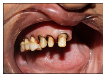
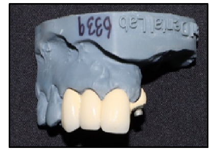
Figure 7
A): Blockout and surveying, B): Designing of framework, C): DMLS metal framework on cast, D): Intraoral framework trial

Figure 8
A): Border moulding and final impression on framework, B): Altered cast, C): Wax rims on altered cast

Wax rims were fabricated using modelling wax. Vertical and centric jaw relation was recorded. Facebow record was made and transferred to semi-adjustable articulator. Teeth arrangement was done (Figure 9 ,Figure 10A). Unilateral balanced occlusion was given on left side and group function on right side. Teeth setting trial was done to evaluate phonetics and esthetics and patient consent was taken.
Figure 9
A): Facebow record, B): Teeth arrangement on semi adjustable articulator, C): Teeth setting trial in patient

Figure 10
A): Final wax up of teeth setting, B): Hollowing of processed denture by removing salt crystals

Flasking of trial denture was done using Type II dental stone (Kalabhai Karson Pvt. Ltd., India) followed by dewaxing. During packing, heat cure acrylic resin (Pink DPI heat cure, The Bombay Burmah Trading Corporation Ltd., India) was used along with cellophane pouch with salt in the defect area was used to create close bulb hollow obturator. Curing was done in an acrylizer unit. Similarly, processing was done for the waxed up mandibular trial denture. After removal of processed maxillary denture, holes were created in the area of defect and midpalate to dissolve salt crystals by running water with the help of syringe (Figure 10B). Cold cure acrylic resin was then used to close the created holes. Female component was picked up in the cast partial denture and nylon cap inserts were placed with the help of mandrel. Finishing and polishing of the prosthesis was done (Figure 11).
Figure 11
A): Intaglio view, B): Frontal view, C): occlusal view of maxillary closed hollow bulb cast partial denture and D): Mandibular denture


Figure 13
Post operative intraoral frontal view, A): Post operative extraoral frontal, B): Left lateral view

During denture insertion (Figure 12,Figure 13), retention, stability, phonetics and esthetics were evaluated. Occlusion was checked in centric, protrusive, left lateral and right lateral excursive movements of mandible. Patient was satisfied with the outcome of the treatment.
Discussion
Various authors like Curtis (1967); Desjardins (1978); Gay & King (1980); Parr et al. (1989) have discussed the framework design for adequate understanding of framework design for different types of maxillary defects. In this case, support was provided by the palate and the remaining natural teeth mostly by the anterior and posterior abutment teeth along with walls of the defect. Since the arch form of remaining teeth is curved, there is scope of tripodal distribution of stresses. Retention was provided with the help of semi precision attachment and direct retainer on first and second molars (embrasure clasp). Indirect retention was provided by the embrasure clasp on premolars and bracing effect of major connector on palatal surfaces of teeth which prevents dislodgement of cast partial denture away from the tissue in the area of defect.
An extra coronal semi precision attachment used in this case was Rhein 83 OT strategy. It has a parallel line support under the ball that automatically aligns the copings, which is important for the insertion of the prosthesis and the durability of the copings, avoiding the risk of ball wear. Hence while fabrication, it was incorporated such that it allowed proper path of insertion and removal of the prosthesis with a passive fit. In this semi precision attachment, male component design is a sphere with a flat head, while the female component consists of retentive nylon caps that are color-coded according to their varying retentive properties. Nylon caps inserts are fitted in the female component of semi precision attachment which enhance retention and stabilization of prosthesis.7 The rationale behind utilization of semi precision attachment was for better stress distribution to remaining anterior teeth by reducing non axial loading and rotational stresses. It also has advantages of better esthetics and splinting of periodontal compromised teeth adjacent to the area of defect with added advantage of better retention, stability and support.8 However, it does have some limitations like being a technique sensitive procedure, adequate understanding of principles and designs should be present. There is a greater need for motivation of the patient for regular follow ups. Repairs and change of retentive nylon caps may be necessary with time.9
Prosthesis in such cases can become heavy thereby causing patient discomfort owing to the large extent of the defect. Various techniques and materials have been reported in literature by Chalian et.al, Brown et.al and Nimonkar et.al. to reduce the weight of the prosthesis. Materials such as silicone, modelling clay, a slurry of plaster and pumice, gelatin, cellophane wrapped in asbestos, salt, polyurethane foam, acrylic resin shim have been used for hollowing the prosthesis. Salt was used in this case due to simplicity of technique along with no residual crystals remaining after curing. It can be easily washed away by creation of hole and running through water.10, 11, 12 Hence reducing the weight of the prosthesis and achieving considerable retention by using semi precision attachment helped meet the needs of the patient.
Conclusion
Combining comprehensive oral examination and devising customized treatment plan along with utilization of recent advancements in fabrication of framework for cast partial denture significantly improves success of prosthesis by improving accuracy, and fit of prosthesis to achieve desired results. It also significantly improves the patient's ability to chew, speak, and swallow, thereby contributing to a better quality of life.