- Visibility 201 Views
- Downloads 7 Downloads
- DOI 10.18231/j.ijohd.2024.030
-
CrossMark

Mastering occlusion: The key to implant success
Introduction
The right occlusal scheme selection for implant restoration is very important, as after osseointegration, excessive mechanical stresses that exceed the physical limits of hard tissues is a major factor in contribution of both initial and long-term bone loss around implants. Occlusal overload can contribute to peri-implant bone loss and lead to the failure of implant prostheses. It is followed by crestal bone loss, thereby deepening the anaerobic sulcus and promoting peri-implant disease. Therefore, it is accurate to state that occlusion plays a crucial role in determining the long-term success of implants. The choice of an occlusal scheme for implant-supported prostheses is broad and frequently debated. Most concepts are adapted from those designed for natural dentition, with some modifications, reflecting similarities in mandibular movement velocity, pattern and muscle usage between patients with implants and those with natural teeth.[1] Additionally, it has been established that achieving clinical success and long-term implant viability requires adherence to biomechanically controlled occlusion. This suggests that the provided occlusion should adhere to sound mechanical principles, primarily directing forces along the implant body's long axis while minimizing off-centered forces. Such an approach should aim to establish and enhance biological stability.[2]
Implant biomechanics
Implant biomechanics is a field that addresses the biomechanical factors potentially detrimental to implants and focuses on modifying these factors to mitigate the cumulative stress that can lead to implant overload. Grasping and controlling these factors is essential for improving the longevity and success of dental implants.
The forces exerted on the implant can be assessed based on their type, direction, magnitude, duration and the presence of any parafunctional forces. According to the basic equation (Stress = Force ÷ Surface Area), it is evident that to reduce stress, either the functional surface area should be increased or the applied force should be decreased.[3]
Numerous factors are considered before fabrication of implant prosthesis. Some are discussed as follows: -
Additional implants are placed in the posterior region as the bite force is more. The anterior teeth can be adjusted to establish proper incisal guidance, thereby preventing posterior interferences during excursions. When natural, healthy canines are present, a canine-guided occlusion is the preferred occlusal scheme for the anterior region. If the canine is absent and has been restored, then a mutually protected occlusion is recommended.[4]
The shape and dimensions of the alveolar arch significantly affect the maximum stress values around the peri-implant bone. The most favourable implant distribution in relation to stress concentration is long ellipsoid narrow shape arch followed by U-shaped long narrow arch, short ellipsoid shape with medium width, U-shaped short and wide and U-shaped medium length and medium width.[5]
Implant-crown ratio- The anatomical ratio refers to the distance from the apex to the shoulder of the implant compared to the distance from the shoulder of the implant to the end of the crown. The clinical ratio is the distance from the apex to the bone level compared to the distance from the bone level to the end of the crown. Clinical crown to implant ratio should not be more than 2.2 as it leads to increased marginal bone loss which may lead to implant failure.[6]
The minimum inter-arch space for a fixed prosthesis is 9-10 mm. Inadequate space can result in structural weakness of the prosthesis, potentially leading to fractures. Consideration should also be given to the stability of the opposing arch and the strength of the patient's muscles. As a general rule, a more rigid opposing arch and greater muscular strength necessitate a stronger implant-supported removable prosthesis, which in turn requires more space.[6]
The position of the smile is notably influenced by the mobility and dimensions of the lips. The position of the tooth from apex to crown is influenced by the extent to which the gingival margin migrates apically after tooth extraction. The critical factor in dental implant placement is the apical-coronal positioning, as errors in this placement can lead to adverse esthetic and biomechanical outcomes. If the dental implant is placed too coronally, the restoration may appear shorter compared to the adjacent tooth. Conversely, if the implant is positioned too apically, significant bone loss can occur, impacting both the proximal bone structure and the facial bone wall height, which can result in undesirable soft tissue contours.[7]
To achieve a proper soft tissue support accurate guided placement of implant placement 3 dimensionally should be achieved and immediate temporization should be done to prevent the collapse of soft tissue. It also reduces the marginal bone loss. If immediate loading is not possible, then adhesive provisional restoration is attached to the transmucosal extension. This establishes the soft tissue support and also helps to preserve inter-arch space.[7]
In cases of bruxism, additional implants are placed to avoid cantilevers and pontics for even load distribution. In clenching, whenever possible, centric vertical contacts should be aligned with the implant's long axis. Narrowing posterior occlusal tables helps prevent unintended lateral forces, reduces the necessary masticatory forces and provides more space for the tongue. Additionally, adjacent implant crowns can be splinted together. Enameloplasty of the cusp tips on opposing natural teeth is recommended to optimize the alignment of vertical forces, following the intended occlusal guidelines. If anatomical limitations preclude placing additional implants in the presence of parafunctional habits, a removable overdenture (RP-4 or RP-5) should be considered. This type of prosthesis can be removed during periods prone to harmful habits.[4]
Balanced occlusion
It is described as the coordinated contact of upper and lower teeth, both on the right and left sides, as well as in the front and back occlusal regions. Balanced occlusion is designed to minimize or restrict the tilting or rotation of denture bases concerning supporting structures. This type of occlusion is not naturally present in dentition. In bilateral balanced occlusion, all teeth make contact during movement, making it particularly useful in the creation of complete dentures.[8]
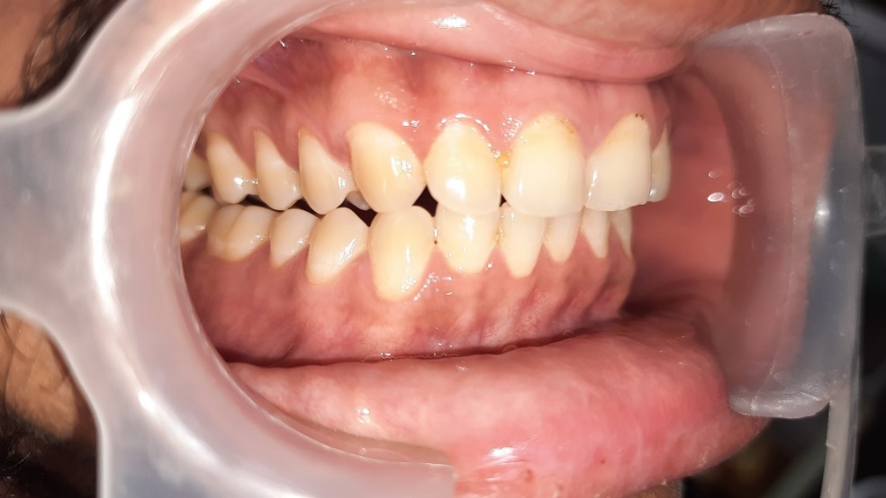
Group function occlusion ([Figure 1])
Unilateral balanced occlusion also termed as group function. It is characterized by the simultaneous contact of the occlusal surfaces on one side of the mouth, facilitating a smooth, continuous glide during lateral movements. In this arrangement, the working side's occlusal load is shared among several teeth, whereas the non-working side remains free of contact, thereby avoiding the imposition of harmful forces on those teeth. This concept was initially identified by Schuyler in 1953.[9]
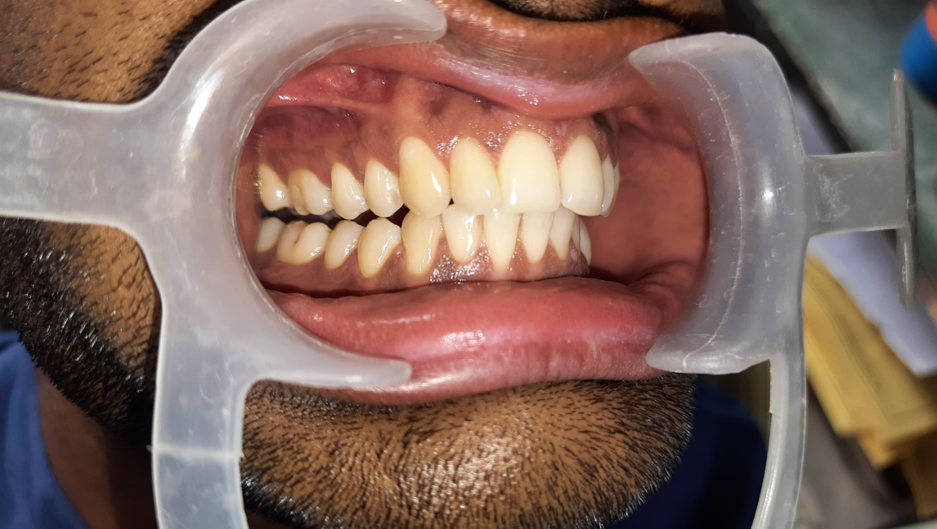
Canine protected occlusion ([Figure 2])
This concept, also referred to as mutually protected occlusion or organic occlusion, involves the upper and lower anterior teeth guiding the lower jaw during lateral or protrusive movements to avoid contact in the posterior region, thus preventing frictional wear. This arrangement is mutually protective: the posterior teeth shield the front teeth when the jaw is in a central position, while the front teeth protect the canines and posterior teeth during protrusion, the canines safeguard the front teeth and posterior teeth during lateral movements.[8]
Implant protected occlusion
Designing an appropriate occlusal scheme is crucial for the longevity of dental prosthetics, particularly in cases of parafunction or poor foundation. A poorly designed occlusal scheme can lead to increased forces on the bone crest, heightening mechanical stress and strain. This can, in turn, elevate the risk of complications that affect both the prosthesis and the surrounding bone support. Carl Misch introduced Implant-Protective Occlusion (IPO), also known as medially positioned-lingualized occlusion, to address and minimize these potential issues.[10]
Discussion
Several elements influence implant protected occlusion and these are discussed as follows.
No premature contacts
Thorough evaluation of premature contacts between maximum intercuspation and centric occlusion is crucial, especially in implant-supported prosthetics. This is because non-mobile implants bear the entire load of the prosthesis when opposed by mobile natural teeth. Consequently, during occlusal adjustments, premature contacts on implants can increase due to potential shifts in the centric position of natural teeth during function.
To determine occlusal adjustments, a thin articulating paper, typically less than 25 μm thick, is used to assess centric relation. This method aims to reduce pressure on the implant crown while increasing contact pressure on adjacent natural teeth. By applying slight occlusal force to the articulating paper, a uniform contact distribution between implant-supported crowns and natural teeth is achieved. It's crucial to note that teeth may take several hours to return to their original positions after enduring significant occlusal forces. Therefore, initially, light forces on adjacent natural teeth are carefully balanced.[11]
Influence of surface area
When the surface area is decreased and the load increases in terms of magnitude, direction, or duration, it leads to increased stress and strain on the interfacial tissue. Several approaches can help address this issue, such as adding more implants in the affected area, performing ridge augmentation, reducing crown height, or widening the implant.[12]
Mutually protected articulation
When natural canines are present, they are crucial during excursions by helping distribute horizontal loads among teeth and enabling the disocclusion of posterior teeth. Additionally, the anterior guidance of implant prostheses with anterior implants should be shallow, as a steeper incisal guidance can increase the force on the anterior implants.[11]
Cusp angle of crown ([Figure 3])
Natural teeth generally have steep cuspal inclinations, while denture teeth are typically designed with a standardized cuspal inclination of around 30%. Cusp inclination is known to produce significant torque, with torque increasing by approximately 30% for every 10-degree rise in cusp inclination.[10]

Implant body angle to occlusal load ([Figure 4])
The direction in which the load is applied, even if it maintains the same force magnitude, can lead to various effects on the bone and implant interface. However, implants are principally designed to endure loads along their long axis. A study carried out by Binderman in 1970 assessed 50 endosteal implant designs and determined that all designs exhibited superior performance under a load along the long axis.[13] Greater angles of load deviation from the implant's long axis led to heightened compressive, tensile and shear stresses, resulting in bone loss and impeding successful bone regrowth.[14]

Crown height
Implant crowns are usually taller than natural anatomical crowns. As the height of the implant crown increases, so does the crestal moment, along with any lateral force components. This amplifies any adverse effects arising from poorly chosen cusp angles, angled implant bodies, or angled loads on the crown due to the heightened crown height.[11]

Cantilever
Cantilevers with an unbalanced crown or implant ratio can amplify the strain on the implant, potentially leading to peri-implant bone loss and prosthesis failure. The stress on the implants increases with the length of the cantilevers and is also influenced by factors such as the number of implants, their spacing and their placement. Lundquist et al.'s clinical study in 1988 demonstrated a link between longer cantilevers and greater crestal bone loss.
A cantilever supported by two implants is a Class 1 lever. When the centers of the implants are 10 mm apart and the cantilever extends 20 mm, a mechanical advantage of 2 is produced. As a result, the load on the cantilever is doubled on the implant farthest from it, while the implant nearest to the cantilever bears the combined stress of both loads.


Occlusal contact position
The quantity of occlusal contacts within a particular occlusal scheme can vary. For instance, according to Peter K. Thomas' occlusal theories, each occluding cusp (stamp cusp), marginal ridge and central fossa should ideally demonstrate a tripod contact pattern, leading to 18 and 15 individual occlusal contacts on mandibular and maxillary molars, respectively. However, alternative occlusal contact schemes may suggest reducing the number of occlusal contacts for molars. Optimal primary occlusal contacts should be situated within the diameter of the implant, particularly within the central fossa, which should measure 2 to 3mm wide in posterior teeth and align parallel to the occlusal plane. Secondary occlusal contacts ought to be positioned within one millimeter of the implant periphery to diminish moment loads, whereas contacts on marginal ridges should be evaded.[15]
Ideal first molar occlusal contacts on natural teeth includes tripod contacts on each buccal cusp, central fossa and marginal ridge.

Implant crown contourIn the upper jaw, the ridge without teeth experiences gradual resorption predominantly towards the inner side, while in the rear part of the lower jaw, resorption tends to happen towards the tongue side. Implants are often centered within the toothless ridge because of the inward resorption, causing them to be placed not directly beneath the outer cusp tip but rather nearer to the central hollow or situated more towards the tongue side, frequently beneath the tongue cusp of the nearby natural tooth. Furthermore, the width of the implant body from cheek to tongue is usually smaller compared to natural teeth.[14]
Occlusal material
Material breakage is very frequent with restorations, whether on natural teeth or implants. Assessing occlusal materials entails taking into account factors such as aesthetics, resistance to impact, management of static loads, efficiency in chewing, susceptibility to breakage and wear, space needed between arches and the positioning of castings.[11]
Design to the weakest arch
In designing implant-supported dental restorations, it's crucial to recognize that failure often occurs at the weakest point within the structure, a principle applicable to any complex engineering system. Therefore, all treatment planning decisions for implant-supported overdentures (IPO) should focus on two primary considerations: first identifying the weakest component in the overall restoration and second developing occlusal and prosthetic strategies to safeguard this vulnerable element. One effective approach to mitigate the force exerted on the system involves incorporating stress-relieving components, which can significantly reduce the impact loads transmitted to the implant support. Consequently, the occlusal scheme may be tailored to favor the complete removable denture recognizing it as the weaker arch in the system.[11]
Timing of loading
Implants can be loaded in different ways like delayed (submerged), progressive bone loading and immediate bone loading. The critical factor influencing the timing of implant placement and prosthetic restoration is bone density.[16]
Conclusion
Dental implants bring an exclusive treatment modality for the restoration of lost teeth. The success of implants is largely controlled by the health of surrounding bone and soft tissue. Excessive stress transmitted to bone-implant interface at the early stage leads to crestal bone loss and early implant failure.
Occlusal considerations in implant dentistry include trans-osteal forces, bone biomechanics, basic biomechanics, differences between natural teeth and implants, masticatory muscle function, occlusal force dynamic and bone resorption. Incorporating these factors leads to the development of an implant prosthesis occlusal scheme.
While natural teeth have a certain level of flexibility that allows them to adapt to occlusal irregularities, implants are less accommodating in this respect. A deficient occlusal arrangement in implant therapy may result in biological and mechanical issues, including implant failure, premature bone loss around the implant, loosening of screws, unseated restorations, fractures in porcelain and peri-implant diseases. Implementing an Implant-Protective Occlusion (IPO) scheme allows the restoration to operate in synergy with the stomatognathic system, thus improving the durability of both implants and prostheses.
Conflict of in Interest
None.
Source of Funding
None.
References
- M Verma, A Nanda, A Sood. Principles of occlusion in implant dentistry. J Int Clin Dent Res Organ 2015. [Google Scholar]
- Y Swaminathan, G Rao. Implant Protected Occlusion. IOSR J Dent Med Sci 2013. [Google Scholar]
- P Prakash, A Narayanan. Biomechanics in Dental Implants. IP Ann Prosthodont Restor Dent 2021. [Google Scholar]
- MS Gowd, T Shankar, R Ranjan, A Singh. Prosthetic consideration in implant-supported prosthesis: A review of literature. J Int Soc Prev Community Dent 2017. [Google Scholar]
- G Sagat, S Yalcin, B A Gultekin, E Mijiritsky. Influence of arch shape and implant position on stress distribution around implants supporting fixed full-arch prosthesis in edentulous maxilla. Implant dent 2010. [Google Scholar]
- AD Fiore, F Maniero, E Stellini. The influence of crown-to-implant ratio on marginal bone loss: a narrative review. Front Oral Maxillofac Med 2020. [Google Scholar]
- N Forna, DA Forna. Esthetic aspects in implant-prosthetic rehabilitation. Med Pharm Rep 2019. [Google Scholar]
- JC Yuan, C Sukotjo. Occlusion for implant-supported fixed dental prostheses in partially edentulous patients: a literature review and current concepts. J Periodontal Implant Sci 2013. [Google Scholar]
- Schuyler CH. Factors of occlusion applicable to restorative dentistry. J Prosthet Dent 1953. [Google Scholar]
- CE Misch. . Dental implant prosthetics 2005. [Google Scholar]
- K Mishra, D Hegde, S Shetty, S Shah, S Sreeram, SL Reddy. Occlusion in implant: At a glance. J Res Dent 2020. [Google Scholar]
- JA Kaukinen, MJ Edge, BR Lang. The influence of occlusal design on simulated masticatory forces transferred to implant-retained prostheses and supporting bone. J Prosthet Dent 1996. [Google Scholar]
- I Binderman, P Shapiro. A modified form for blade type endosteal implant. Oral Implantol 1972. [Google Scholar]
- MW Bidez, CE Misch. Force transfer in implant dentistry: basic concepts and principles. J Oral Implantol 1992. [Google Scholar]
- PK Thomas. . Syllabus on Full Mouth Waxing Technique for Rehabilitation Tooth-to-tooth Cusp-fossa Concept of Organic Occlusion 1967. [Google Scholar]
- GI Benic, JM Mari, CH Hämmerle. Loading protocols for single-implant crowns: a systematic review and meta-analysis. Int J Oral Maxillofac Implants 2014. [Google Scholar]
- Introduction
- Implant biomechanics
- Balanced occlusion
- Group function occlusion ([Figure 1])
- Canine protected occlusion ([Figure 2])
- Implant protected occlusion
- Discussion
- No premature contacts
- Influence of surface area
- Mutually protected articulation
- Cusp angle of crown ([Figure 3])
- Implant body angle to occlusal load ([Figure 4])
- Crown height
- Cantilever
- Occlusal contact position
- Occlusal material
- Design to the weakest arch
- Timing of loading
- Conclusion
- Conflict of in Interest
- Source of Funding