Introduction
Teeth that are not part of the typical series of deciduous or permanent dentition are referred to as supernumerary teeth. They can appear in any part of the mouth. They can appear as a single tooth or multiple teeth, unilaterally or bilaterally, erupted or impacted, in the mandible/maxilla or both jaws, and unilaterally or bilaterally. Supernumerary teeth are more prevalent in the permanent dentition, with a prevalence of 0.1 to 3.8 percent.1 The upper maxilla is more likely than the mandible to have supernumerary teeth.2 Although the basic cause of supernumerary teeth is unknown, various explanations have been presented, the most popular of which is that these teeth grow as a result of horizontal dental lamina proliferation or hyperactivity. Supernumerary teeth can occur anywhere in the dental arch, however they are most commonly found on the maxillary midline between two maxillary central teeth, which are known as mesiodens. The maxillary molar, maxillary lateral incisor, mandibular premolar, and mandibular molar are usually next. Conical, tuberculate, supplementary, and odontomatous teeth are morphologically characterised according to their shape.3 Supernumerary teeth can lead to displacement, crowding, root resorption, dilaceration, loss of vitality of adjacent teeth, delayed eruption or failure of eruption of permanent teeth, subacute pericoronitis, gingival inflammation, periodontal abscesses, dental caries due to plaque retention in inaccessible areas, incomplete space closure during orthodontic treatment, and pathological problems such as dentigerous cyst formation, ameloblastomas, odontomas, and fistulae.1
Case Report
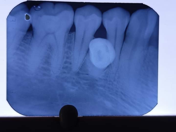
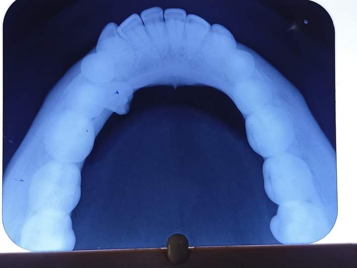
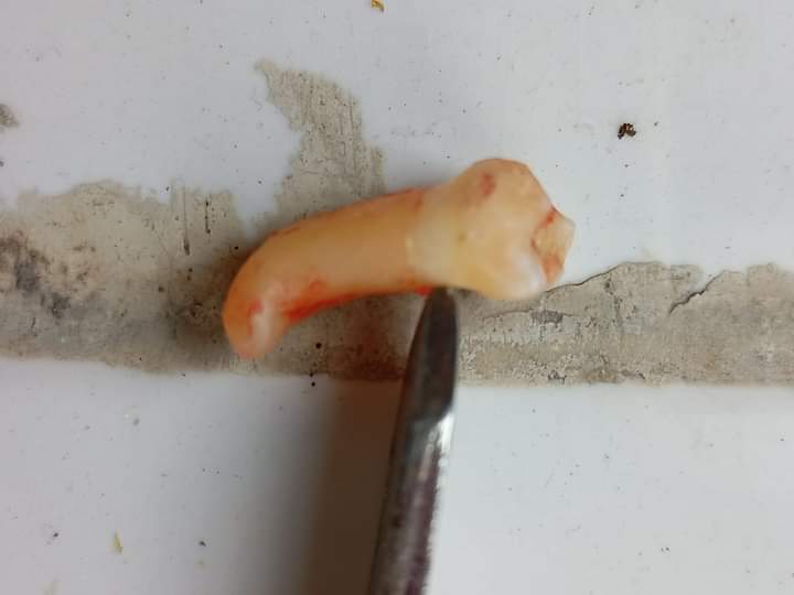
A 33 year old male reported to the Department of Oral Medicine And Radiology in Sri Sukhmani Dental College and Hospital, Dera Bassi, Punjab, India, with a chief complaint of irregularly placed tooth in the lower right back region of the mouth since the last 5 years. Medical and family history was non-contributory. There were no abnormalities found during the extra-oral clinical evaluation. On intra oral examination, a supernumerary tooth lingual to first and second right mandibular premolars was seen and it was completely erupted in the oral cavity. The cuspal anatomy of the tooth resembled the Mandibular premolar. Clinically, it had pierced through the lingual alveolar ridge. The supernumerary tooth was extracted under local anaesthesia, because it was causing irritation to the tongue.
Discussion
Supernumerary teeth in the premolar area, unlike other supernumeraries, are more common in the mandible. They can appear singly or in multiples, be erupted or impacted, but the majority are found to be asymptomatic and unerupted.4 In the present case, however it was single, asymptomatic and fully erupted in the oral cavity. It was discovered that non-syndrome multiple supernumerary teeth were more likely to appear in the mandible. When several supernumerary teeth were studied by particular sites for both jaws, the premolar area was the most common, followed by the molar and anterior regions, respectively.5 In the literature, dentigerous cyst and root resorption have been mentioned as common complications linked with supernumerary premolars. In order to avoid complications such as damage to the mental nerve and blood vessels, clinicians should be cautious when removing supernumerary premolars surgically. Because recurrence of supernumerary premolars following surgical removal has been recorded, proper follow-up using panoramic radiographs is particularly crucial.6 In this case, it was irritative to the tongue, so it was extracted under local anaesthesia. A abrupt change in the axial inclination of the root or between the crown and the root of a tooth is known as dilaceration. Root dilaceration is a rare developmental abnormality that usually affects the posterior teeth.7 Dilaceration of the tooth can be caused by a number of different circumstances. Mechanical stress to the primary precursor tooth is the most frequently known cause of tooth dilation. Other probable causes that have been identified in the literature include primary tooth germ developmental anomalies, advanced root canal infections, ectopic tooth germ development, lack of space, and inherited factors.8 In this case, dilacerations of root is seen in supernumerary tooth.


